Mar 14, 2019

Incontinence for Stroke and Other TBIs: Well, SOMEBODY Had to Write About It and It Might As Well Be Me

Once upon a time, before I had the stroke, I used to sit for long, agonizing hours at my office desk, and didn't urinate for 12 hours while drinking boatloads of coffee and water. I didn't want to leave my desk for fear the boss wanted something earth-shattering, silly office things, like telling us who passed gas in front of the Director or who got canned because he watched Westerns on his office computer. He always wanted us available for things. Things that could wait

As a result, I got used to not peeing. I developed a remarkably humongous bladder (5 doctors at 5 offices in 3 different states said so after I had CT scans) and I had, I might add, constant UTIs (urinary tract infections). You're supposed to "pee" after drinking massive amounts of fluid.


Anyway, despite my inordinately large bladder, I developed incontinence after I had my stroke. I was constantly leaking. So I took to Poise to help with the embarrassment, larger and thicker pads until I was at #6, the Ultimate. Ultimate absorbency, the ominous sign above a grocery shelf said. Ultimate absorbency. I had reached the limit. 


Why was this happening? Soon, in about a week's time of research, I had my answers. And we're off!


No matter how you explain it, everything comes from the brain. And The American Urological Association (AUA) has a simple answer. 


It's called a neurogenic bladder, or bladder dysfunction. 


"The bladder and kidneys are part of the urinary system," the AUA says. "These are the organs that make, store, and pass urine. When the urinary system is working well, the kidneys make urine and move it into the bladder. The bladder is a balloon-shaped organ that serves as a storage unit for urine. It is held in place by pelvic muscles in the lower part of your belly."


The AUA goes on to say that the nerve signals in your brain let you realize that your bladder has to empty itself. Then the  brain tells the bladder muscles to contract, allowing urine out through your urethra, the tube that carries urine out of your body. Your urethra muscles are called sphincters that keeps the urethra shut until you're ready to "pee." 


If these nerves are damaged by illness or injury, the muscles may not be able to relax or tighten at the correct time. As a result, bladder muscles may be overactive and squeeze more often than normal before the bladder is full, or sometimes the muscles are too relaxed and let urine come out before you're ready, or sometimes the sphincter muscles around the urethra remain tight when you are trying to empty, and sometimes people have both overactive and underactive bladder at different times. Don't bother with the distinctions. If you're leaking or gushing, you're wet to some degree. 
Maybe you're a visual learner, so how about this? Remember. Start at the top with the brain.


If you have neurogenic bladder, or incontinence, see your doctor. It can't be cured, but it can be managed. 

I came across 2 interesting therapies, the one involving surgery, the other a needle:


Sacral Neuromodulation: When drugs or lifestyle changes don't help, there's sacral neuromodulation. The sacral nerves carry signals between your spinal cord and the bladder, allowing the surgeon to place a narrow wire in proximity to the sacral nerves. A wire is connected to a small, battery- operated device that is placed under your skin. The harmless electrical impulses to the bladder stop the signals that can cause the bladder to leak.


Percutaneous Tibial Nerve Stimulation: This type of  neuromodulation involves a needle that's inserted into a tibial nerve in your leg, most likely the ankle. The needle, connected to a device that emits electrical impulses, travel to the tibial nerve, and then to the sacral nerve. This procedure is done in your doctor'ss office, and patients ordinarily receive 12 treatments for top results. 


The AUA says that certain drinks, foods, and medications may act as diuretics, stimulating your bladder to "go" more often. They include:

  • Alcohol
  • Caffeine
  • Chocolate
  • Carbonated drinks and sparkling water
  • Heart and blood pressure medications, sedatives, and muscle relaxants
  • Large doses of vitamin C


Persistent urinary incontinence may be caused by underlying changes, including:
  • Neurological disorders, like stroke or other TBIs 
  • Pregnancy
  • Childbirth
  • Age changes
  • Menopause
  • Hysterectomy
  • Enlarged prostate
  • Prostate cancer
  • Obstruction, such as a tumor or urinary stones
  • Hysterical laughing or annoying coughing


Risk factors that increase your risk of developing urinary incontinence include: 
  • Gender
  • Age that weakens the muscles involved with urination
  • Being overweight
  • Brain injury
  • Smoking
  • Family history (lousy genes will get you every time)
  • Other neurological diseases 
  • Diabetes

As I said before, urinary incontinence may not be preventable, but you have to maintain a healthy lifestyle, including: 
  • Maintain a gender-specific correct weight
  • Practice pelvic floor exercises 
  • Avoid bladder bothers listed above 
  • Don't smoke, the perennial favorite
  • Avoid constipation by eating more fiber, constipation being one of the causes of urinary incontinence
Easier said than done? Maybe. But as the quote-worthy Mark Twain once said, "The only way to keep your health is to eat what you don't want, drink what you don't like, and do what you'd rather not." 

I think Mark Twain nailed it.

Mar 1, 2019

Having a Stroke Is Easy: It's What Comes After, aka Post-Stroke Psychiatric Syndromes

Ok. So you had a stroke, and now the drama is over? Wrong! As I always say, having a stroke is the easy part compared to what comes after. There are 7 conditions named here.

In an article in Psychiatric Times, the drama is simply unfolding. 

The researchers say the most commonly reported of these in the studies are:


1. post-stroke depression (called PSD as opposed to PTSD ((Post Traumatic Stress Disorder)) which ALL stroke survivors have) and 

2. post-stroke dementia (PSDem), which may affect mood and cognitive symptoms. The researchers say that assessment of the survivor's living situation, level of support, and attention to caregivers' and family members' behavioral observations are necessary with patients who have cognitive impairment or other neurologic impediments to communication. 

A higher risk of PSD has been found in patients with "left-sided cortical and basal ganglia lesions and to lesions closer to the frontal lobe than to left posterior or right frontal lesions." (This is why I say--all the time--to get as much information from your doctors as possible in regard to what exactly happened to your brain). 


PSD may involve a spectrum of mood disorders,  "vascular depression," and dementia-related depression. (Vascular depression often predating stroke is associated with higher degrees of cognitive impairment). Because these relationships between depression and dementia are currently vague, the physician should offer treatment of PSD early in the post-stroke period to strengthen mood and cognitive function.


Most reviews have found the risk of PSD to be between 20% and 79% for up to 18 months post-stroke, and the risk of depression is nearly twice that in persons who have not had a stroke. Untreated patients are at risk for chronic illness (even after 2 years) although patients with PSD may recover spontaneously within 12 months with tricyclic antidepressants (TCAs), selective serotonin reuptake inhibitors (SSRIs), and psychostimulants (methylphenidate).


PSDem symptoms include poor social judgment, amnesia, cognitive disorganization, language deficits, apraxia, disinhibited and/or unsafe behavior, paranoia, and visual-spatial dysfunction. Thoughts of suicide have been reported up to 24 months after stroke. Patients with PSDem should be followed up monthly, with reassessment of cognitive examination, repeated depression inventory, and screening for psychotic symptoms.


3. Post-stroke mania is rare, but it may be associated with right-sided stroke. Manic symptoms include decreased need for sleep, expansive and/or irritable mood, increased goal-directed activity, disregard for social constraints, recklessness, talkativeness, racing thoughts, excessive laughter or giggling, and poor judgment. Persons with post-stroke mania should routinely be referred for psychiatric care.

4. Anxiety symptoms include episodes of panic, excessive sweating, worrying, and decreased sleep. Risks of 26% for men and 39% for women have been found in a post-stroke anxiety disorders study. A combination of anxiety and depression may be more usual in left cortical stroke, while depression without anxiety may be more usual in left subcortical stroke.

Here's a tip: Avoidance of benzodiazepines is important. These agents may cause cognitive decline, verging on PSDem. Follow-ups should be done in monthly increments. 


Less frequently seen post-stroke psychiatric symptoms include pathologic crying and pathologic laughter (PBA), apathy, and isolated fatigue. 


5. Pathologic laughter and crying are sometimes grouped as pathologic emotions (PE, another name for PBA) with sudden bursts of either laughter or crying, despite of the mood state. 

6. Apathy in the absence of depression may be difficult to evaluate, but it's a complete lack of initiative without tearfulness, sleep/appetite disturbance, hopelessness, or suicidality.

The use of psychostimulants for PSD or apathy, sometimes both, may be associated with a risk of seizure and/or cardiac side effects. Concern for these risks must be balanced against undertreatment of PSD. In cases of cardiac disease, consultation with a cardiologist is best with careful dosing of psychostimulants.

7. Post-stroke psychotic disorder symptoms include delusions, hallucinations, thought disorganization, and regressed motor behavior. Post-stroke psychotic disorder has been shown with right-sided lesions and cortical/subcortical atrophy. Persons with post-stroke psychosis should be referred for psychiatric care. A patient with pre-stroke psychotic illness, such as schizophrenia, may ultimately decline following a major stroke.

Having a stroke is the easy part. It's what comes after that's usually a bitch. 

Feb 8, 2019

Why You Lose Memory from Brain Injury and Possible Ways to Improve It

This post is for everyone because you never know when you’ll suffer a brain injury. I had a stroke. And with that may come memory loss. I want to say I learn something new every day. In reality, I don't, but pretty close to it. Researching and completing this post over a 2-week period taught me many things about memory, and I'll share them with you. 


Memory is defined as: the faculty of the brain by which information is encoded, stored, and retrieved when needed, vital to experiences, and is the retention of information over time for the purpose of influencing future action. 


Let's get academic for the moment if we haven’t already. This from LumenLearning:
  • It is theorized that memories are stored in neural networks in various parts of the brain associated with different types of memory, including short-term memory, sensory memory, and long-term memory.
  • Memory traces, or engrams, are physical neural changes associated with memories. Scientists have gained knowledge about these neuronal codes from studies on neuroplasticity.
  • Encoding of episodic memory involves lasting changes in molecular structures, which alter communication between neurons. Recent functional-imaging studies have detected working-memory signals in the medial temporal lobe and the prefrontal cortex.
  • Both the frontal lobe and prefrontal cortex are associated with long- and short-term memory, suggesting a strong link between these two types of memory. Damage there is monumental.
  • The hippocampus is integral in consolidating memories but does not seem to store memories itself.
For all those who know what part of the brain-sustained damage, and you should demand to know by asking your doctor, the following descriptions of the different types of memory will be useful to determine where your loss is according to where it's positioned. 

image

  • Sensory Memory: 
Brain part most relevant: Temporal and  Occipital lobes 
The ability to retain impressions of sensory information after the original stimuli have concluded and t
he shortest-term element of memory through the five senses of sight, hearing, smell, taste and touch, which are retained accurately, but very briefly. 

  • Short-Term Memory:
Brain part most relevant: Pre-Frontal Cortex, Frontal, and Parietal lobes 
Short-term memory is followed hand-in-hand with sensory memory and allows the ability to hold on to a piece of information temporarily in order to complete a task or remember directions, for example.

Long-Term Memory:
Brain part: Frontal and Temporal lobes 
Long-term memory allows the ability to decode information, create associations among an object’s various properties, and develop opinions.


(For Sensory and Long-Term Memory: The Temporal lobe is where lies the hippocampus, a small organ which forms an important part of the limbic system, the region that regulates emotions and events that happened in the past. Needless to say, it's complicated).

And this from McGill U in Canada:
In regard to long-term memory, researchers found two types: 

Declarative memory, aka explicit memory, is what happens when you recall your birthday or what you recall eating last night. You can name and describe each of these remembered things explicitly.


Non-declarative memory, aka implicit memory, is like riding a bike or tying your shoelaces, where you do things, most often motor skills, that do not require the use of language.  

Here's the bottom line: sensory memory leads to short-term memory which leads to long-term memory. If anything is going on where the process is interrupted, you will have some sort of memory issue.

Another study by researchers at the University of Queensland in Australia found out that, relative to short term memory, using magnetic resonance imaging (MRI) machines, people perform tasks requiring them to hold information, such as the location of a flash of light, and the PFC (pre-frontal cortex) becomes active. In terms of the sides of the brain, the left is more involved in verbal memory while the right is more involved in spatial memory, such as recalling where the flash of light happened. 

So can you do anything to get your memory restored? It depends. The amount of damage is certainly important.  

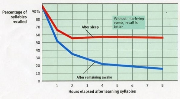
One study said practicing meditation, drinking coffee (albeit before 2pm if you have trouble sleeping at night), eating berries (blueberries are high in flavanoids), chewing gum (because it may improve concentration), and doing exercise (the brain is stimulated) can help improve memory. But the most important? Getting enough sleep.

A study reported on memory- challenged participants to memorize illustrated cards, testing their memory. Then they had a 40-minute break--one group napped and the other stayed awake. After the break, both groups were tested on their memory recall of the cards. Surprisingly, the group who had napped performed better, retaining on average 85 percent of the patterns compared to 60 percent for those who had remained awake.


On the other hand, the brain is the least understood part of the human body. But practicing meditation, drinking coffee, eating berries, chewing gum, doing exercise, and getting enough sleep are harmless, and one or some of those activities may do the trick. 

You know what I always say: if it works, use it.